SMON: a case study
An Odyssey through germ theory – part IV
If you haven’t read the previous article in this series, it’s advisable to do so here.
SMON (‘subacute myelo-optic neuropathy’), is a ‘disease’, who’s defining manifestation was an epidemic in Japan that began at the end of the 1950’s, and lasted until approximately 1970.
The first two cases were identified in 1958 and were presented in a paper titled: “A cured case of hemorrhagic diarrhea accompanying polyneuritic syndrome”. 4 more cases were identified in 1960, and two more in 1961. Thereafter, “similar reports followed one after another, and it became evident in 1963 or so that there was a sharp increase in sporadic cases and some localized outbreaks” (Kono, 1971).
Overall, estimates place the number of people affected at around 10,000, but according to the College of Medicine at the University of Tokyo, this figure could be as high as 30,000.
Geographically, it seems to have touched most areas of Japan, although for reasons we will come back to, it is noteworthy that “severe outbreaks of SMON had occurred in two rural areas in Okayama Prefecture since 1967” (Kono, 1971).
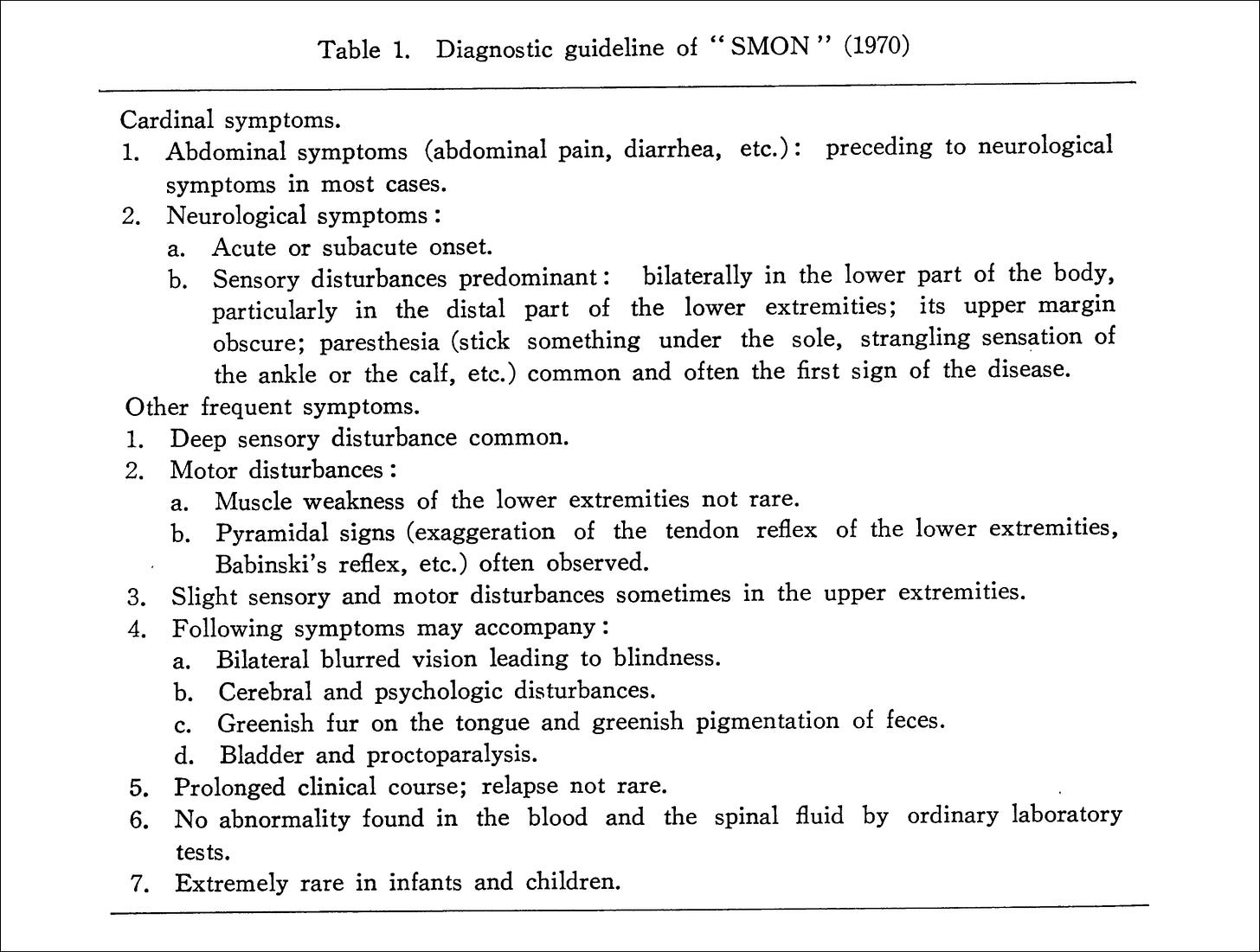
The ‘disease’ was described as follows: “in the end of 1950's Japanese physicians noticed an occurrence of a myelitis-like illness preceded by abdominal symptoms, such as abdominal pain or diarrhea or both, before the appearance of neurological disorders…
At the beginning, the disease was suspected to be akin to benign encephalomyelitis, similar to Iceland disease (White and Burtch, 1954) or a special type of multiple sclerosis accompanying abdominal symptoms, and was taken by physicians as Devic's disease in some outbreaks.
However, soon these concepts were abandoned when the clinical analysis proceeded and the accumulated evidences of post-mortem findings uncovered a unique feature of the disease” (Kono, 1971).
Although the ‘disease’ was said to be remarkably similar to others (including ‘multiple sclerosis’ – which we’ll come back to), we are told it had a ‘unique feature’ and that “accordingly, it was once called ‘Nonspecific encephalomyelitis with abdominal symptoms’” (Kono, 1971).
What is this ‘unique feature’? It would appear to be the “accompanying abdominal symptoms”. Various sources have stated that this was deemed to be of ‘cardinal importance’, but that subsequently, “less importance was attached to this aspect” (Baumgartner et al., 1979). Some, however, have stated that “subacute combined degeneration (SCD) of the cord is the main neurological differential diagnosis of SMON” (Meade, 1975).
It is interesting to note that despite the use of the word ‘optic’, in all cases “total blindness was found in 2.5 % and blurred vision in 18.9 %” (Kono, 1971). Also noteworthy is that “the disease develops often in hospitalized patients with other chronic illnesses, such as malignant neoplasms, chronic nephritis, tuberculosis, diabetes etc” (Kono, 1971).
Was SMON really a new ‘disease’? This question was asked right from the start; “a question arises, however, whether or not the disease constitutes a distinctive clinicopathological entity, since it is well documented that a variety of neurological disorders may be accompanied by acute abdominal conditions” (Tsubaki et al. 1965).
And although it was said that “neurologists are in general agreement that SMON exhibiting all its neurological features … was a distinct and real clinical syndrome”, the ‘incomplete form’ was said to account for most cases, rendering it “not easily distinguishable from other causes of peripheral neuropathy and myelopathy” (Meade, 1975).
Such was the confusion that researchers in Bombay, looking at the incidence of SMON in India, asked; “how does one diagnose SMON, especially in its incomplete form, in the absence of any specific laboratory tests”?
Another interesting point to note – which curiously doesn’t feature in the ‘differential diagnosis’ – is that “Takasu, Igata and Toyokura (1970) noticed that the fur of the tongue was coloured green in some SMON patients” (Kono, 1971). Green faeces and urine were also found in certain cases (Igata, 2010).

It is also noteworthy that the “incidence maximum” was during the summer. A similar pattern was seen in the case of ‘polio’ in the United States and Northern Europe. ‘Polio’ and other ‘diseases’ that ‘mimic it’, is particularly relevant to this discussion, given that, flaccid paralysis is said to be one of the symptoms of SMON – which is also said to be the ‘unique feature’ of ‘polio’. And as discussed in the previous article, these symptoms are in fact, not unique to ‘polio’, but are shared by a vast range of conditions said to be caused by all manner of ‘viruses’ and bacteria.
So, what was the cause of this new ‘disease’? Several avenues were explored, including that of a novel ‘virus’ – and this, appears to have been the preferred theory: “for virtually the whole course of the epidemic, which ran from 1955 to 1970, SMON was regarded as probably being an infectious disease” (Meade, 1975). And because of this, the Japanese government “considered patient isolation. The victims experienced crude discrimination, resulting in a series of suicides … the commonest cause of death among victims was suicide” (Imamura, 2007).
In the previous article, we outlined the three overarching steps virologists must complete in order to demonstrate they have found a novel ‘virus’.
We will now examine each of these in turn.
Isolation
A ‘virus’ – initially called ‘SMON virus’ was ‘isolated’ by Inoue et al. in 1970; “virus was isolated, with a high frequency, in BAT-6 [hamster tumour] cell cultures accompanying a weak and incomplete cytopathic effect (CPE) from feces and spinal fluid of SMON patients”.
The ‘cytopathic effect’ is one of the key markers virologists look for when ‘isolating’ a new ‘virus’. The ‘isolate’ is added to a cell culture preparation – a petri dish containing cells coming from a variety of sources. If cell death is subsequently observed, this is interpreted as (indirect) evidence that the particle in question is the cause of the ‘disease’ being investigated.
In this case, the ‘isolate’ consisted of fluids extracted from faecal matter and spinal fluid samples taken from those suffering from the affliction. These were then added to various cell culture preparations, made from monkey kidney cells, human embryonic kidney cells, and BAT-6 (hamster tumour) cells.
At this stage, we can already see a problem with this method – namely, that no ‘isolate’ was used in the cell culture, and therefore, the ‘isolate’ can create the desired effect for reasons other than the presence of a ‘virus’. For instance, in this textbook on poliovirus, the authors write: “virus culture of stool specimens presents challenges due to toxicity and an abundance of other microorganisms.” Additionally; “the primary inoculation of the specimen extract will enable virus attachment … but this can occur in competition with cell degeneration due to material present in specimen extracts that are toxic to the cells.”
Other fluids can also be problematic – in this textbook on influenza, the author states that “occasionally, some samples (especially oral fluids) may cause cytotoxicity, which can mimic viral CPE to some degree.”
Isolation is defined as; “to set apart from others”. It is clear that no such thing has happened here – if it had, the issues highlighted above would not be a problem. And this situation is not unique to the case at hand. For instance, with Sars-Cov-2, we are told that the ‘virus’ was ‘isolated’ in the following way; “the patient's oropharyngeal samples were obtained by using UTM™ kit … we inoculated monolayers of Vero cells with the samples and cultured the cells at 37°C in a 5% carbon dioxide atmosphere.”
In their paper, Inoue and his colleagues stated that a “weak and incomplete” cytopathic effect was observed. The following images are provided as evidence of this.
The first image shows ‘BAT-6’ (hamster tumour cells) that were inoculated with the ‘uninfected materials’. This preparation was photographed ‘unstained’. The second image shows the same thing – but of cells to which ‘infected materials’ had been added. Presumably, these were subsequently ‘stained’.
‘Staining’ refers to the application of various (toxic) heavy metals (mostly lead, but also osmium and uranium) to the sample in order to allow the specimen to be viewed more clearly. How does this impact the experiment?
This methodology – that of ‘cultivating viruses’ in cell lines, was originally devised by John Enders and colleagues, for which they were awarded the Nobel prize. In their experiments, they used the ‘measles virus’ in much the same way as what is described above. When describing their own results, they stated that “a second agent was obtained from an uninoculated culture of monkey kidney cells. The cytopathic changes it induced in the unstained preparations could not be distinguished with confidence from the viruses isolated from measles.”
In other words, they carried out a control experiment using ‘uninfected’ materials, and what they observed – in their own words – was indistinguishable from that which they observed in the experiment that used ‘infected’ materials. They then wrote: “but, when the cells from infected cultures were fixed and stained, their effect could be easily distinguished since the internuclear changes typical of the measles agents were not observed.”
At first, it would appear that they carried out the exact same procedure with both ‘infected’ and ‘uninfected’ materials – and the difference was only noticed after ‘fixing and staining’. But just like the Inoue paper we are looking at, no mention is made of ‘fixing and staining’ the sample taken from the ‘uninfected’ culture – suggesting that they only followed these steps for the ‘infected’ culture – and therefore are not comparing like for like.
This point was raised with a virologist on Twitter. The individual posed the challenge of finding another paper that attempted to replicate Enders’ findings, but no such paper could be find – which is curious, given that we’re told attempts are usually made to reproduce such findings.
Characterisation
Building on the aforementioned research, another paper published in 1970 sought to provide further information about the new particle: “recently, Inoue observed cytopathogenic effect of millipore-filtered extract of patient's feces on culture cells and considered this as the successful isolation of the causative virus of SMON.”
The researchers carried out the following procedure: “some of the culture media for virus propagation contained a small amount of tetracycline. For electron microscopic observation, cells showing cytopathogenic effect 4 to 6 days after the virus infection were collected, fixed with glutaraldehyde and osmium tetroxide and embedded in Epon. The sections were observed by the electron microscope after uranyl acetate and lead stainings.”
‘Fixing’ refers to the use of various chemicals, including some of the aforementioned, and others, such as glutaraldehyde, which are added to the sample in order to ‘halt’ any metabolic processes, and preserve the sample in a state that is as close as possible to what it would have looked like whilst alive. So it’s worth noting at this stage that anything scientists are looking at under their electron microscopes is not alive; they have never observed phenomenon such as ‘viral replication’ in real-time.
The researchers write that: “in addition to the above observation, numerous mycoplasma ranging from 150 mIl. to 800 mp. in diameter were observed together with the virus particles in the specimens prepared from tetracycline-free cultures”.
‘Mycoplasma’ are a type of bacteria, said to be ‘disease-causing’, and have been found to be associated with all manner of ‘diseases’, including; “respiratory infections, urogenital infections, fatiguing illnesses, autoimmune diseases, neurodegenerative and neurobehavioral diseases and complications affecting the central nervous system, cardiac infections, oral infections, peridontal diseases, sexually transmitted diseases and systemic infections found in various solid cancers and leukemias and immunosuppressive diseases, such as HIV-AIDS” (Nicolson, 2019).
This list also includes cases of ‘polio’ and so-called ‘Guillain-barré syndrome’ (which itself is said to ‘mimic polio’). Note that the words ‘caused by’ and ‘associated with’ appear to be used interchangeably. And if you recall, in the previous article, we found that another ‘disease-causing bacteria’ – streptococci – was found to correlate positively with exposure to arsenic.
A control experiment was carried out, and they wrote that “the virus particles and mycoplasma were not observed by the electron microscope in the control cells of the BAT 6 cell line.” So, it would appear that the procedure itself hasn’t induced the changes.
The authors also let us know that “the virus particles and mycoplasma were not observed by the electron microscope in the control cells of the BAT 6 cell line.” In other words, they ran a control experiment and here, they found no ‘virus’. For whatever reason, no imagery is provided comparing the two samples, so we’ll just have to trust that this was indeed the case.
They conclude their study with the following: “the first successful electron-microscopic observation of a virus isolated from a patient with SMON was performed. The morphological and developmental characteristics of this virus suggests that this type of virus has not been isolated from humans. Hence, it is considered that the virus observed is of a new type and presumably the causative agent of SMON.”
And they also note that: “the virus particle observed in the present study closely resembles mouse leukemia virus in the morphological aspect. However, the followings are the characteristics of the present virus different from leukemia virus. The formation process of this virus virion is much more similar to Japanese encephalitis virus.”
To summarise; it would appear that after adding the ‘infected’ materials to the cell cultures, a particle was found in the culture that had been inoculated with the ‘infected’ materials, but not in the culture that was ‘uninoculated’. ‘Disease-causing’ bacteria were also found, but for whatever reason, these appear to not be relevant.
Establishing causation
In 1972, Inoue et al. published another paper where they attempted to use their isolated virus to produce the disease in mice. In these experiments, the virus “was inoculated intracerebrally or intraperitoneally into C57BL/6 newborn mice at birth.”
‘Intracerebral’ and ‘intraperitoneal’ inoculations refer to injections of the materials into the brains and stomachs of the test subjects. And this stage, you might be asking yourself; why are injections needed when ‘viruses’ are usually said to spread through aerosol or skin contact?
The experiment appears to have yielded the expected results; “the diseased mice had paralysis of the legs after an incubation period of 2-3 weeks or more.” Furthermore, it would appear that the virus was not recovered from the brains of the mice used as controls (who were either not inoculated, or inoculated using uninfected materials); “the virus was detected in high titre from the brains of the diseased mice, whereas it was not recoverable from the brain of one control mouse which showed wasting and ruffling of hair.”
The experiment done here is in no way unique – this is standard procedure, and the practice appears to go all the way back to Robert Koch’s anthrax experiments. We previously discussed Thomas Rivers’s paper on ‘chickenpox’. In this study, he did the following:
“Blood was drawn from patients with chicken-pox usually during the first 24 hours after the appearance of the eruption. The blood was not citrated and before clotting occurred was injected in 2 cc. amounts into each testicle of normal rabbits (1,800 gm.)."
At the time of inoculation the needle was moved about in the tissues to produce a certain amount of trauma ... 4 days later the testicles were removed, ground up thoroughly with sterile, chemically clean sand, and mixed with 10 cc. of physiological salt solution … Then 1 cc. of the emulsion was injected into each testicle of normal rabbits. Two areas on the rabbits' skin were shaved and scarified. One of the areas was smeared with the emulsion, the other was used as a control. An eye of each rabbit was also inoculated.”
When confronted on this issue, virologists and others will often say that such papers are old, and thus are no longer relevant (although as demonstrated in the above screenshots from Twitter, the relevance seems to be entirely arbitrary – the age of the paper is only ever brought up when convenient). The monkeypox article was due to be published in a major alternative online publication, but the editor decided not to proceed with publication, after one of their scientific advisors brought up the age of the paper as a point of contention.
So, the same virologist from Twitter provided a video of ‘viruses’ moving around in cells. When asked how he knows what these particles actually do, he shared a paper from 2012, where they apparently attempted to transmit the ‘vaccinia virus’ to mice (‘vaccinia’ is closely related to the ‘smallpox virus’, ‘variola’. The former was used to produce ‘smallpox vaccines’). In this paper, the methodology involved shaving some mice, anesthetising them, and then “applying 1 × 10⁷ plaque-forming units of VV [vaccinia virus] to the dorsal skin and scratching 20 times with a bifurcated needle”.
The end result was this:
He, and others was then asked why no controls were used in this experiment, and whether we would not expect to see similar lesions on any animal subjected to such a procedure (note also how these lesions look nothing like what ‘smallpox’ is said to look like in humans).
Success?
So what’s happened so far? Our researchers have, apparently completed all of the above steps. In theory then, they have discovered a new ‘virus’ and proven that it causes the ‘disease’ being investigated.
However, the authorities later confirmed that the SMON epidemic was in fact, caused by the anti-diarrheal drug clioquinol. The drug was banned in Japan, and various other countries followed suit.
In a letter published in The Lancet, 1971, Professor Tsubaki wrote that: “we have observed a strong association between this drug [clioquinol] and severe neurological disturbances … we found that 166 (96%) of 171 patients with S.M.O.N. had taken clioquinol, mainly for digestive disorders, before the onset of their neurological symptoms …
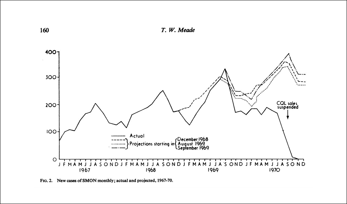
It was also confirmed that the increase in the occurrence of S.M.O.N. in Japan was very closely connected with the increased production and sales of clioquinol …
Due to these findings, the Ministry of Health and Welfare prohibited, in September, 1970, the production and selling of clioquinol in Japan. Since then we have heard of very few patients developing S.M.O.N.”
So as we can see, all the ‘ingredients’ are there. Every box was ticked. The only problem; we are told no ‘virus’ was to blame; the ‘disease’ was the end-product of exposure to a toxin – in this case a drug.
It is also most interesting to note that other ‘viruses’ were discovered in patient materials:
“Several investigators isolated CPE agents from the spinal cord, spinal fluid, blood and feces. They were echovirus type 2, coxsackie A group viruses, herpes-like new virus, enterovirus of unknown type (Nakazawa et al., 1968, 1969) etc. However, the etiological role of all these isolates has not been confirmed by other investigators. Another suspect is Mycoplasma which was isolated from the fur of the tongue and feces …
In other patients higher antibody titers against EB [epstein-barr] and Marek's disease viruses were demonstrated. One investigator reported a higher incidence of complement fixing antibody against the Mycoplasma pneumoniae antigen…
We do not know what these results imply” (Kono, 1971).
All of this leaves us with some important questions; what then, was the particle the researchers discovered? Why were so many different ‘disease-causing viruses’ found in the materials taken from those affected by the ‘disease’. Why did they observe a ‘cytopathic effect’ in cell culture? Why did the mice who were part of the control group, not get sick?
In short; why were they able to tick all the boxes that would theoretically have demonstrated the existence of a new, ‘disease-causing pathogen’, but then decide that the new ‘pathogen’ had nothing to do with any of it?
Furthermore; why were (most) of the patients taking clioquinol in the first place? And why is it that SMON has never been reported in countries outside of Japan, where the drug was more widely used? Was clioquinol used as a decoy?
In the next article, we’ll take a closer look at clioquinol, and what might in fact be, the real cause of the phenomenon discussed here.
References
Baumgartner et al. (1979) – Neurotoxicity of halogenated hydroxyquinolines: clinical analysis of cases reported outside Japan
Domenico et al. (2012) – Susceptibility to Vaccinia Virus Infection and Spread in Mice Is Determined by Age at Infection, Allergen Sensitization and Mast Cell Status
Enders et al. (1954) – Propagation in Tissue Cultures of Cytopathogenic Agents from Patients with Measles
Igata (2010) – Clinical studies on rising and re-rising neurological diseases in Japan – A personal contribution
Imamura et al. (2007) – History of public health crises in Japan
Inoue et al. (1971) – Virus Isolated from Patients of Subacute Myelo-Optico-Neuropathy (SMON) in Japan
Kono (1971) – Subacute Myelo-optico-neuropathy, a new neurological disease prevailing in Japan.
Martín (2016) – Poliovirus: Methods and Protocols
Meade (1975) – Subacute myelo-optic neuropathy and clioquinol. An epidemiological case-history for diagnosis
Nicolson (2019) – Pathogenic Mycoplasma Infections in Chronic Illnesses: General Considerations in Selecting Conventional and Integrative Treaments
Ota (1970) – Electron microscopic demonstration of a new virus isolated from a patient with SMON
Park et al. (2020) – Virus Isolation from the First Patient with SARS-CoV-2 in Korea
Rivers (1923) – Studies on varicella
Spackman (2020) – Animal Influenza Virus: Methods and Protocols
Tsubaki et al. (1965) – Subacute Myelo-Optico-Neuropathy Following Abdominal Symptoms: A Clinical and Pathological Study
Wadia (1977) – Some observations on SMON from Bombay


Another (2) superb articles from you and Caroline. In Awe of you both! Thank you so much.
Just mentioned and linked to this wonderful article in my new interview ... Exposing the Many Fallacies of Today’s “Science” w/ Dr. Jordan Grant https://www.bitchute.com/video/9bdHKhbQlxrK Dr. Jordan, a board certified urologist, has been highly critical of the virology methodologies used to justify the COVID-19 fiasco, going so far as to question, with great acumen and insight, the very existence of those allegedly disease-causing vectors known as viruses.