Disclaimer: nothing in this article is to be taken as medical advice. The information provided here is to be used purely for informational and educational purposes.
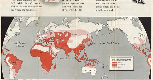
Malaria is defined by the World Health Organisation (WHO) as a “life-threatening disease caused by parasites that are transmitted to people through the bites of infected female Anopheles mosquitoes”. According to the latest World Malaria Report published by the WHO in 2021, in 2020 there were an estimated 241 million malaria cases in 85 countries, 95% of which were reported from Africa. More than 50% of global malaria infections are reported from five countries: Nigeria, Congo, Uganda, Mozambique and Angola. In 2020, there were 627,000 death reported, 96% of which were reported from these five countries.
The word ‘malaria’ – or ‘mal’aria’ (‘bad air’) is a term said to have originated in Italy and was introduced into the English language in 1827 by Scottish geologist John MacCullogh, “as a substitute for the more restricted ‘marsh miasma’ or ‘paludal poison’”. Although malaria is now said to be a problem primarily confined to the southern hemisphere, and in particular the African continent, it was once endemic in Europe, having been introduced into the continent as a result of “favorable geomorphological and climatic conditions” and “the presence of adequately sized human and competent vector populations”. Taking the example of Italy; at the end of the 19th century, the total number of malaria deaths ranged from 15,000 to 20,000 per year. Italy was declared ‘malaria-free’ by the World Health Organisation in 1970.
In the UK, where some say that it was once referred to as ‘marsh fever’ or ‘ague’, medical historian Dr Mary Dobson writes that malaria “had a striking impact on regional patterns of disease and death”. By the end of the 19th century, ‘indigenous malaria’ had “clinically disappeared from England”, and according to Dobson, one possible reason for this is “as housing and ventilation improved, there was probably a reduction of summer temperatures at the more influential micro-level, hindering the course of infection in the domestic mosquitoes”. Today, “deaths from malaria have continued to occur, but have been from imported cases”. Dobson writes; “few doctors in Britain are acquainted with this “tropical” disease, and several cases of imported malaria have recently been misdiagnosed”.
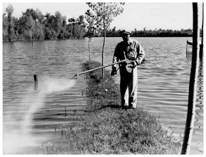
In the US, malaria was “considered eliminated” by 1951, whilst in Europe, this feat was accomplished in 1974, thanks to “man-made contraction of vector breeding sites and improvement of living standards” and “widespread drug treatment and residual insecticide spraying”. One such insecticide was Paris Green; a highly toxic compound made of copper and arsenic which we have already discussed in some detail here. In the US, Paris Green was first used to control the spread of the Colorado potato beetle; “in the summer of 1867, farmers in Illinois and Indiana applied Paris green in a desperate attempt to destroy the destructive insect. Word of this efficacious insecticide spread rapidly. Within the first decade of its introduction as an insecticide, in excess of 500 tons of Paris Green were sold annually in the New York City market alone. And London Purple (approximately calcium arsenite), a by-product of the aniline dye industry, entered the market as a rival insecticide”. It is interesting to note that the following year, “readers of the Veterinarian, an English journal, were informed … that a ‘very subtle and terribly fatal disease’ had broken out among cattle in Illinois. The disease killed quickly and was reported to be ‘fatal in every instance’”. This disease, now known as ‘Texas fever’ was eventually said to have been caused by a protozoan known as Pyrosoma bigeminum, spread by cattle ticks.
A few decades later, in 1921 “a United States Public Health Service officer showed that it was effective as a mosquito larvicide when sprayed on water.” Up until that point, oil had been used; it was “spread on larvae-infested water as a means of making it nearly impossible for the larvae to breathe”. Poisoning using Paris Green however, was found to be far more effective; “the mosquito larva, which lives in pooled, stagnant, or slow-moving water, must come to the surface to poke its air tube out of the water to breathe, even though it feeds below the surface. This makes it vulnerable to a poison such as Paris Green that, when mixed with a light inert powder, floats on the surface of water”.
DDT (‘dichlorodiphenyltrichloroethane’) was also used extensively. It was first created in 1874 by Austrian chemist Othmar Zeidler. According to an article in the New Yorker he “threw it away”, having apparently, not “realised its value”. It was ‘rediscovered’ as an insecticide by Swiss chemist Paul Müller in 1939, for which he was awarded the Nobel Prize in 1948.
The initiative to use these compounds for ‘disease control’ appears to have been spearheaded by the International Health Board (1913 – 1928) and the Rockefeller Foundation (1913 – present). Their work was carried on by the Office of Malaria Control in War Areas (the CDCs predecessor), which was established in 1942 to “limit the impact of malaria and other vector-borne diseases (such as murine typhus) during World War II around military training bases in the southern United States and its territories”.
Indeed, both malaria and typhus (which we’ll come on to shortly) were major problems during both world war I and II. In Europe, 1944, “nearly 900,000 pounds of 25 percent Paris Green in lime and 7,000 gallons of oil (mostly No. 2 diesel with varying contents of DDT) were applied by airplane for American and British malaria control. During 1945, about 250,000 pounds of Paris Green mixed with diatomaceous earth (1: 3 for American use) or cement (1: 6 for British use) and roughly 72,000 gallons of 5 percent DDT in oil were dispersed by plane in the theater”.
Discovering plasmodium
A French army doctor, Charles Louis Alphonse Laveran (1845-1922) first observed the malaria parasite in 1880; “while looking through a crude microscope at the blood of a febrile soldier, he saw crescent-shaped bodies that were nearly transparent except for one small dot of pigment”. This brownish-black pigment – known as ‘hemozoin’ or ‘malaria toxin’ is the “product of hemoglobin digestion by the malaria parasite”. Prior to Laveran, hemozoin “had been found in cadaveric spleens and blood of malaria victims by several investigators including Meckel, Virchow, and Frerichs”. Laveran subsequently examined blood samples from 192 malaria patients but only found the same ‘crescents’ in 148 of them. He was awarded the Nobel Prize for this discovery in 1907.
In 1897, Surgeon-Major Ronald Ross (1857-1932) of the British Indian Medical Service “discovered a clear, circular body containing malarial pigment in a dapple-winged Anopheles mosquito that had previously fed on an infected patient”. The following day he repeated the experiment and “observed even larger pigment-containing bodies”, after which “he published his observations” in the British Medical Journal. Ross too, was awarded the Nobel Prize. In 1948, Henry Shortt and Cyril Garnham discovered how the parasites first develop in the liver before spreading to the blood, after experimentally infecting human volunteers with P. vivax (unfortunately, we were not able able to locate the paper where the procedure they used was documented).
The parasite in question – known as ‘plasmodium’ is what is commonly referred to as a ‘protozoan’; a single-celled eukaryote (an organism whose cells contain a nucleus). According to the CDC, there are approximately 156 species, and of these, four are said to cause ‘disease’ in humans; P. falciparum, P. vivax, P. ovale and P. malariae. Other varieties are implicated in the infection of other animal species; for instance, rodents are said to be infected by the likes of P. berghei, P. yoelii, and P. chabaudi.
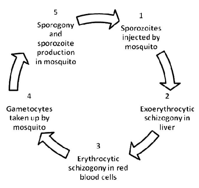
The infection cycle can be summarised as follows. During a blood meal, an infected female Anopheles mosquito inoculates ‘sporozoites’ into the host. These are carried around the body until they eventually reach the liver where they ‘infect’ liver cells and undergo a phase of asexual multiplication (‘exoerythrocytic schizogony’), and eventually mature into ‘schizonts’. These ‘schizonts’ rupture, releasing ‘merozoites’ that travel through the body, infecting ‘erythrocytes’ (red blood cells) along the way. This is known as the ‘blood-stage’ of infection, and it is the parasites in this form that are said to be responsible for the clinical manifestations of the disease. Once inside the blood cells, they initiate a second phase of asexual multiplication (‘erythrocytic schizogony’), which results in the production of about 8-16 more merozoites that go on to invade new red blood cells.
As the infection progresses, some young merozoites develop into male and female ‘gametocytes’ that circulate in the peripheral blood. Eventually some are ‘sucked up’ when a mosquito has a blood meal. Once inside the mosquito, the ‘gametocytes’ mature into male and female ‘gametes’, after which fertilisation occurs and an ‘ookinete’ is formed within the mosquito’s gut. This ‘ookinete’ penetrates the gut wall and becomes an ‘oocyst’ within which another phase of multiplication occurs resulting in the formation of sporozoites that eventually migrate to the mosquito’s salivary glands. When the mosquito next has a blood meal, the sporozoites are once again injected into the host, and the cycle repeats.
A unique ‘disease’?
According to the CDC, symptoms usually begin 10 days to 4 weeks after infection, although this can occur up to a year later. However, like other ‘diseases’ said to be caused by various microorganisms, infected hosts are often asymptomatic, suggesting that the parasite alone is not usually sufficient to cause the ‘disease’ in question. According to a paper published in March 2022, “the current prevalence of asymptomatic malaria in children living in the southwest of the Central African Republic is very high”. A literature review published in 2012 notes that “despite a wealth of studies on the clinical severity of disease, asymptomatic malaria infections are still poorly understood” and that “asymptomatic malaria remains a challenge for malaria control programs as it significantly influences transmission dynamics”.
When a given individual does exhibit symptoms, these, according to the CDC, can include generic symptoms described as “fever and flu-like illness, including shaking chills, headache, muscle aches, and tiredness”. Nausea, vomiting, and diarrhoea can also occur, and in some cases anaemia and jaundice. If left untreated, “the infection can become severe and may cause kidney failure, seizures, mental confusion, coma, and death”.
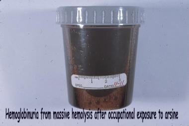
Another symptom of ‘malaria’ is referred to as ‘blackwater fever’, also referred to as ‘hemoglobinuric fever’, which consists “of a destruction of red blood cells so widespread that the liver, being powerless to transform the liberated hemoglobin into bile pigment, the greater part is excreted by the kidneys”. Interestingly, this paper, published in 1907, noted that “the parasitic findings in hemoglobinuric fever are at great variance. A Plehn states that he seldom found them soon after the outbreak and only exceptionally later; F. Plehn found no parasites; Marchiafava and Bignami report them absent in numerous cases; Koch detected parasites in 2 out of 16 cases; Sambon says that they are absent in a considerable proportion of cases; Nocht found them in 75 per cent, of cases; Cardamatis in 4 of 25 cases; Ollwig in 6 of 15 cases; Crosse pronounces them frequent; Bertrand found them in almost all cases; Shropshire in 41 per cent, of cases …” and so on.
‘Blackwater’ urine is also one of the symptoms of exposure to arsine gas.
These symptoms are also shared with a number of other ‘diseases’ said to be caused by pathogens spread by ‘arthropods’ (invertebrate animals with an exoskeleton, such as mosquitoes, spiders, mites and ticks). These include bacteria, ‘viruses’, and protozoa. ‘Arbovirus’ is a ‘portmanteau word’ used to describe any ‘virus’ transmitted in this way. ‘Co-infection’ is also common.
Examples of ‘diseases’ said to be caused by a ‘virus’ transmitted in this way, and / or said to be readily confused confused for malaria, include Yellow fever (“a more severe case can be confused with severe malaria”), Ebola (“symptoms of Ebola virus infection and malaria overlap to a great extent”), West Nile virus, Japanese Encephalitis virus (“we report a case of concurrent infection of JE and mixed plasmodium infection”), Dengue fever (“there was significant number of misdiagnosed cases of DV for either malaria or typhoid”), Crimean-Congo Hemorrhagic fever (“Hemorrhagic manifestations can occur in this disease due to thrombocytopenia or DIC the same as can be observed in malaria”), Lassa fever (“Lassa fever presents with symptoms and signs indistinguishable from those of febrile illnesses such as malaria and other viral haemorrhagic fevers such as Ebola”), Rift Valley virus (“the symptoms of RVF mimic many other endemic infections in these areas, such as malaria”), Zika (“the first human ZIKV isolate came from a 10-year-old Nigerian female in 1954. ZIKV was isolated in mice inoculated with the patient's serum. Interpretation of the clinical presentation of the patient was difficult because the patient's blood also contained numerous malaria parasites”) and Chikunguya (“in Ethiopia, febrile patients are often misdiagnosed with malaria or typhoid fever”).
Examples of ‘diseases’ said to be caused by bacteria that are transmitted in this way include typhus and typhoid (“both typhoid and malaria share rather similar symptomatology and epidemiology. Malaysia is endemic for both these diseases and one should not be too surprised when faced with a diagnosis of co-infection of typhoid and malaria, as have been described in India and Canada”), Rocky Mountain Spotted Fever (originally known as ‘black measles’), Leptospirosis (“the clinical signs and symptoms of uncomplicated malaria and leptospirosis were similar, making accurate clinical diagnosis difficult without laboratory confirmation”) and Q Fever.
These diseases also often mimic conditions said to be caused by other ‘viruses’, such as ‘polio’, which also mimics a number of other ‘diseases’, various ‘auto-immune diseases’, and even those said to be caused by ‘nutritional deficiencies’ such as ‘scurvy’. For instance, in the case of Zika, “clinical evidence suggests that Zika virus contributes to Guillain-Barré syndrome that causes temporary paralysis”. Zika is said to cause ‘microcephaly’ (a birth defect in which a baby's head is smaller than expected) in the offspring of women who are infected during pregnancy, however this has also been observed in cases of malaria.
‘Cerebral malaria’ is said to be “most severe pathology caused by the malaria parasite”, and may be diagnosed as ‘Guillain-Barré syndrome’. Interestingly the linked case report notes that “patient 1 progressed to develop respiratory paralysis and required mechanical ventilation”. A WHO report published in 2022 documenting the progression of “a vaccine-derived poliovirus type 2 epidemic” in Ghana, states that “malaria was the most probable cause [of] about 39% of AFP [acute flaccid paralysis] cases”. In the case of typhus, a report published in 2022 documents a case of “Scrub Typhus Presenting as Acute Flaccid Paralysis in a Child”. The child was treated with doxycycline (an iron chelator), and her symptoms gradually improved.
Like bacteria and fungi, plasmodium is iron-hungry; “iron is an essential micronutrient required by all living organisms including malaria parasites for many biochemical reactions, especially growth and multiplication processes”. It therefore needs to take up iron from either inside and / or outside the parasitised red blood cells. ‘Hemolysis’ refers to red blood cell death (lysis), at which point their contents are released into the surrounding blood plasma. As discussed here, this can occur for any number of reasons, including exposure to various poisons such as arsenic. Free heme, which is released when this occurs, is toxic to cells, so the parasites convert it into an insoluble crystalline form called ‘hemozoin’ – the ‘malaria pigment’ Laveran and others observed.
‘Hemolytic anemia’ occurs when bioavailable iron drops, as a result of too much hemolysis. Because of this, malaria is said to be a “major cause of anemia in endemic areas” – however it does “not cause iron deficiency, but iron deficiency does reduce the incidence of severe malaria … iron deficiency and malaria still often coincide in the same patient”.
It is also interesting to note that malaria is said to be one of the ‘opportunistic infections’ that affects HIV patients (and the symptoms of ‘primary HIV infection’ are “often mistaken for malaria”). As discussed here, ‘immunosuppression’ is said to be either ‘primary’ or ‘secondary’. ‘Secondary’ refers to ‘immunosuppression’ said to be caused by ‘environmental factors’ which according to the British Society for Immunology can include; “HIV, malnutrition, or medical treatment (e.g. chemotherapy)”. No mention is made of exposure to ‘pollutants’ such as arsenic, although the ‘immunosuppressive’ effect of it is well documented.
Establishing causation
The presence of the parasite in diseased patients does not automatically mean that they are the cause of a given disease. As we previously discussed here, Koch’s postulates were developed to provide a framework so that scientists could demonstrate causation when investigating the alleged pathogenicity of a given microorganism.
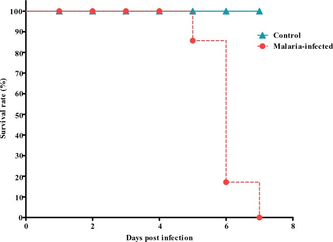
So called ‘human models’ are seldom used, however various ‘animal models’ have been produced over the years. For instance, in a paper published in 2012, experimenters sought to induce ‘cerebral malaria’ in mice. The method they used involved injecting infected red blood cells into the test subjects ‘intraperitoneally’ (into the abdomen). Following inoculation, the ‘passaging’ technique was employed, whereby the blood of an infected animal is injected into another, and this procedure repeated several times. Controls received an equal amount of uninfected blood cells.
Deleterious effects were observed in the mice inoculated with the infected cells; “20% mortality was recorded on day 5 post infection, followed by 70% mortality on day 6 and by day 7, 100% mortality were observed”.
Two things are of note here. First, as seems to so often be the case in these experiments, the alleged pathogen does not appear to have been isolated (separated from all else). Instead, contaminated red blood cells were injected, which raises the question of whether the blood may be contaminated with anything else. If, for instance, the presence of the parasite is a marker for exposure to one or more environmental pollutants – such as lead, arsenic or cadmium (which we will discuss shortly), then clearly the presence of such pollutants would have an impact on the test subjects.
The original method for ‘ex vivo’ (out of body) cultivation of plasmodium was devised in 1977 by James Jensen and William Trager. Their process involved adding nutrients to the culture – blood in this case – several times a week. The same process seems to carry on today. According to a manual published in 2010 by the WorldWide Antimalarial Resistance Network “the culture medium has to be changed every 24 hours in a sterile environment”. Human blood appears to be widely used, however a paper published in 2002 notes that “for reasons that include cost, reproducibility, and possible presence of inhibitory immune factors and antimalarial drugs, there is interest in substituting other types of mammalian sera (bovine, monkey, horse, goat, sheep, rabbit, or swine) for human serum or even developing a serum-free medium for parasite cultivation”. The mention made here of antimalarial drugs and ‘inhibitory immune factors’ being a problem is particularly interesting as this clearly demonstrates that there is an issue with contamination. Astonishingly, however, antimalarial drugs appear to be the only concern and it is unclear what kind of toxicological analysis is performed (if any) when blood is ‘harvested’ for use in these cultures. If environmental pollutants are indeed an issue, then all of the aforementioned animals are likely to be affected, and this can be more or less of a problem, depending on the species. For instance, in 2004, researchers found that “15-30-fold more arsenic species were bound to the Hb [haemaglobin] of rat RBC [red blood cells] than that of human RBC”. Other sources of contamination include animal feed. A study published in 2015 found that “all diets were contaminated with pesticides, heavy metals (mostly lead and cadmium), PCDD/Fs and PCBs”, leading the researchers to conclude that “the chronic consumption of these diets can be considered at risk”.
Second, it appears to be increasingly acknowledged that certain ‘parasite-host’ interactions can, in fact, be beneficial. An interesting case in point comes from a study published in 2007; “Parasites as heavy metal bioindicators in the shark Carcharhinus dussumieri from the Persian Gulf”. In this paper, the authors sought to investigate “lead and cadmium concentrations in the liver, intestine, muscle and gonad of the shark Carcharhinus dussumieri and its parasites, Anthobothrium sp. and Paraorigmatobothrium sp. (Cestoda)”. According to the researchers, “the results strongly support the view that helminth parasites are extremely sensitive early warning bioindicators, particularly in sensitive environments under threat but where pollution levels are presently low. They may also have a beneficial effect on the health of their hosts by acting as heavy metal filters”.
There seems to be some evidence that plasmodium may, in some cases at least, play a similar role. Austrian physician Julius Wagner-Jauregg (1857-1940), won the Nobel prize for medicine in 1927 “for his discovery of the therapeutic value of malaria inoculation in the treatment of dementia paralytica”.
Exploring the toxic route
Francesco I de' Medici (1541–1587) was the second Grand Duke of Tuscany. He died suddenly, along with his wife, Bianca Capello. “Tertian malarial fever” was reported as the cause of death. At the time, rumours spread that the couple had in fact been poisoned by Francesco's brother, Cardinal Ferdinando. In 2006, a research team carried out a toxicological analysis of his remains, and concluded that the results of their analysis were “consistent with the hypothesis that the Grand Duke and his wife were victims of an acute arsenic poisoning”.
Then, in 2010, Fornaciari et al. found evidence of the presence of P. falciparum in the skeletal remains of Francesco I de’ Medi, leading the authors to conclude that malaria was indeed the cause of his death. These findings, according to them, “absolve Ferdinando I from the shameful allegation of being the murderer of his brother and sister-in-law”.
In a letter published in 2015, Donatella Lippi, PhD writes; “information recently discovered in the Vatican Library now adds support to the conclusion of poisoning … Francesco's official autopsy reported postmortem signs of arsenic poisoning, such as velvety red congestion of the stomach, and an unofficial report by doctors who witnessed the autopsies of Francesco and his wife refers to a “poison which had corrupted their internal organs.” Lippi concludes; “the presence in human remains of arsenic concentrations at lethal levels does reliably identify acute arsenic poisoning as the cause of death. Autopsy descriptions and the Vatican document also support this conclusion”.
Arsenic is not the only substance that seems to be associated with the presence of plasmodium. Several studies have examined the relationship between blood-lead levels (BLL) and the incidence of malaria. According to a literature review published in 2021, “BLL associated with decreased risk of malaria was demonstrated by two studies conducted in Benin and Nigeria, while BLL associated with increased risk of malaria was demonstrated by a study conducted in Nigeria. BLL was associated with the risk of severe malaria, involving severe neurological features and severe anemia”. Similar results have been observed in the case of mercury exposure in the Brazilian Amazon; “the odds of reporting a past malaria infection was four times higher for those also reporting a history of working with mercury”.
In a paper published in 2008 investigating “Lead poisoning associated with malaria in children of urban areas of Nigeria”, the authors write that “interest in malaria-lead poisoning interaction has been remarkable in being non-existent”. This study, according to the authors, “is one of the first to find a significant negative association between BLL and malaria in a pediatric population, and this association remained significant after controlling for confounding diseases and symptoms “. They go on to say that “the suppression of malaria symptoms by lead is not a new observation, however. From the Middle Ages until recent times, Fowler’s solution and other lead compounds were used extensively in the treatment of malaria, although the efficacy and benefit of such treatment are now questionable … With lead poisoning, over 99% of blood lead is intra-erythrocytic and during anemia, lead becomes even more concentrated in red blood cells … It seems reasonable to suggest that increased concentration of lead in erythrocytes may inhibit the development from the ring form via the trophozoite to the schizont stage following Plasmodia infection, especially since these life stages must feed on the lead-enriched erythrocyte”.
The authors then state that “toxificication of the food source (erythrocyte) can be considered a possible mechanism by which lead exposure reduces the parasitemia in Plasmodium-infected individuals. High intraerythrocyte concentration of lead can affect several essential cellular processes adversely, including inhibition of protein synthesis leading to disruption of proper iron utilization by Plasmodia in ways that can exacerbate iron deficiency”.
The authors also gently remind us that “some people may claim that lead poisoning is an antidote for malaria – nothing can be further from the truth”. Indeed, what this study seems to suggest is that some level of lead poisoning may result in a malaria infection. Too much however, and the parasite perishes.
Poisons everywhere
Both lead and arsenic poisoning are a widespread problem. According to a report published by UNICEF in 2020; “1 in 3 children – up to 800 million globally – have blood lead levels at or above 5 micrograms per deciliter (µg/dL), the level at which requires action. Nearly half of these children live in South Asia”.
To give just one example; Kabwe (Zambia) was home to a lead mine from 1904-1994. According to Human Rights Watch, during that period, “smelter fumes covered much of the surrounding soil with lead dust. Since then, seasonal flooding and windblown dust from the mine dump, as well as ongoing small-scale mining, have worsened the contamination”.
It is interesting to note that contaminants such as these are spread in much the same way that ‘viruses’, bacteria, protozoa and other microorganisms are said to. Going back to the aforementioned paper published in 2008 that investigated lead poisoning amongst children in Nigera, the authors write that “after deposition, some of the contaminated dusts are cycled back into the air, as evidenced by the trailing plume of dusts that accompany lorries in many parts of Nigerian cities. The lead contaminated dusts become pervasively redistributed from the roadside to peoples’ homes and yards and play areas where they come into contact with children”. They also note that “the length of time a child plays outside can be regarded as a surrogate for duration of contact with contaminated dusts, hence the significant association with BLL. Although boys are generally allowed to play outside more often, this does not translate into a significant BLL–gender relationship. Girls tend to stay at home with their mothers and are likely to be more exposed to household-related risks (such as house dusts and indoor air pollution) which may mitigate the effects of playground exposure for boys.” Interestingly, the authors also write that “pet ownership is found to be a predictor of childhood BLL because their furs serve as moving collectors of lead dust which can be transferred to human hosts; in Nigeria, pets are rarely groomed by their owners”.
Arsenic contamination is also widespread in Africa. A paper published in 2015 sought to “build a map of arsenic distribution in African waters using existing data and to highlight the lack of data comparing to the probable magnitude of the problem”. In this document, all values of arsenic in water greater than 10 μg L−1 were considered high. Arsenic concentrations in groundwater ranged between 0.02 and 1760 μg L−1 , and up to 10,000 μg L−1 for surface water. Most of the countries for which data was available recorded values considered high (> 50 μg L−1). These included Morocco, Burkino Faso, Ghana, Togo, Nigeria, Ethiopia, Tanania, Zimbabwe, Botswana and South Africa. Malaria is said to be a risk in all these countries, with the exception of Morocco. However, a paper published in 2015 notes that “typhoid fever is a major public health problem in Meknes city (Morocco)”.
According to the authors of this pan-African study “these high levels of arsenic in the surface water are usually related to the mining operation … indeed, areas devoid of mining activities like Okavango Delta have generally low values of arsenic in the surface water, whereas in mining activity areas the surface waters have high levels of arsenic”. However, in some cases, such as Rift Valley (an area located in East Africa that spreads over seven countries), high levels of arsenic in the surface water were found, but the area is said to be devoid of any mining activities. Other researchers have highlighted that “high concentrations (6460 μg L−1 ) of arsenic in the surface water in the vicinity of Lomé and other big cities … was due to the impact of the effluents from the industrial activity as well as hazardous waste dumping”.
Going back to Europe, we previously discussed the pervasiveness of arsenic in everyday life. Lead too, was a problem; “the citizens of Ulm in Germany were plagued by agonising stomach cramps in the 1690s. But it was soon noted at a local monastery that some of the monks, who happened to abstain from drinking the popular local wine, were being spared by God. The source was eventually identified as a lead oxide sweetener added to the wine - and then eliminated via what was possibly the world's first formal ban on the use of lead. In England, these same stomach cramps became known as ‘Devon colic’ after a similar 17th Century outbreak, this time caused by the lead used in local cider presses”.
It is sometimes said that so-called ‘marsh gas’ was the cause of ‘malaria’ in places like England. ‘Marsh gas’ – or methane, is “commonly found in aquifers contaminated with arsenic”, and the researchers found in their study that “methanotrophic bacteria used organic carbon in methane as an electron donor to reductively dissolve the minerals, freeing sequestered arsenic”.
Contaminated water, contaminated insects
Various studies have sought to examine how metal exposure impacts insects. One such study published in 2018 sought to understand the effect of metal pollution on insecticide resistance of Anopheles mosquitoes. According to the authors, “metal pollution is one of the most important anthropogenic pollutants Anopheles mosquitoes are exposed to, both in urban and rural areas”. In this study, they found that these mosquitoes and their larvae demonstrated resistance; “these data suggest an enzyme-mediated positive link between tolerance to metal pollutants and insecticide resistance in adult mosquitoes. Furthermore, exposure of larvae to metal pollutants may have operational consequences under an insecticide-based vector control scenario by increasing the expression of insecticide resistance in adults”.
As of late, insects are increasingly being promoted as an alternative source of protein. In 2016, a study investigated any risk factors relating by studying the potential for bioaccumulation of cadmium, lead and arsenic in larvae of two insect species, Tenebrio molitor (yellow mealworm) and Hermetia illucens (black soldier fly), both of which are said to be a potential future food source. The researchers found that bioaccumulation “was seen in all treatments (including two controls) for lead and cadmium in black soldier fly larvae, and for the three arsenic treatments in the yellow mealworm larvae”.
Other studies have found similar results with other insect species, such as beetles; “the beetles from polluted areas accumulated extremely high amounts of As in their bodies”. Another study published in 2013 sought to understand the ‘trophic transfer’ (transfer through the food chain) of arsenic from aquatic insects to terrestrial predators. They found that in certain species, trophic transfer does occur, although in others environmental exposure plays a more important role. Bioaccumulation appeared to occur in all species tested.
In 2014, researchers studying the accumulation of lead, cadmium, mercury, and chromium, in adult cockroaches found that “both males and females underwent metal accumulation”. In 2013, a paper sought to investigate the effect of arsenic exposure to Culex mosquitoes, said to be the vector for transmission of ‘West Nile Virus’, ‘Japanese encephalitis virus’, ‘St Louis virus’ and others. The researchers state that their results “indicate tolerance of these Culex species to arsenic exposures”, and that “there was also significantly more arsenic accumulated … when exposed to arsenate than arsenite”.
In 2020, a paper investigating the “effect of heavy metals accumulation on locomotor activity of Ixodid ticks”, considered “pathogenic of many human diseases”, found that heavy metals like cadmium and lead bioaccumulate.
In 2021, a study investigating the “possibility of using an arsenic (As) hyperaccumulator as a bioinsecticide”, and its effect on hawk moths found that arsenic bioaccumulates in the larva of the species; “the As concentration in the larva body was as high as 850 mg kg−1. Such high concentration of As in the larva body might have been the case that T. clotho lacks a process to exclude As”.
Mosquitoes have a long mouthpart (‘proboscis’) that is used to piece the skin and suck blood out. When this happens, saliva is secreted. In a study published in 2018, researchers note that mosquito saliva is a “complex mixture of proteins” that has “profound effects on the human immune system”. It “allows the mosquito to acquire a blood meal from its host … by circumventing vasoconstriction, platelet aggregation, coagulation, and inflammation or hemostasis”. More than a hundred proteins are involved and many “have unknown functions”.
These can include ‘Anophieline Antiplatelet Protein’ (AAPP), ‘Aegyptin’, ‘Hamadarin’ and ‘Heparin’. Some of these, such as the anti-coagulant AAPP contain a number of cysteine-residues. Cysteine contains sulphur, which arsenic binds with readily, as Caroline discusses in more detail here.
Summary
Malaria, like other ‘diseases’ that we have already covered in some detail – including the various ‘poxes’, does not appear to be a unique condition. The symptoms are shared with a great many other ‘diseases’ said to be caused by a variety of ‘viruses’, bacteria and protozoa, all of which share symptomology with various forms of poisoning, arsenic in particular. In the case of plasmodium, it is not entirely clear that this microorganism is the fundamental cause of the problem. Indeed, the existence of ‘asymptomatic carriers’, suggests other variables play a part – such as exposure to certain environmental pollutants, who’s symptoms also just so happen to be shared by almost every ‘disease’ we’ve looked at.
It is interesting to note that in the case of ‘blackwater fever’, a number of examples were provided of people suffering from the condition, but where plasmodium was absent in either some, or all cases. This was also the case when Laveran’s first observed plasmodium in some malaria patients. Like the studies involving ‘viruses’ and bacteria, there are a number of issues with the ‘animal models’ involved in demonstrating that the organism in question is the cause of the disease, and not merely associated with it. The flaws in the experimental methodology provide an important clue as to what the problem may in fact be, and the findings presented here suggest the aforementioned pollutants seem to spread in the exact same way that the ‘disease-causing’ that are allegedly the source of the problem, do.
Another great mystery lies in the question of why the ‘vectors’ involved in the spread of these microorganisms – mosquitoes in particular – appear to be aware of geopolitical borders. Malaria for instance is said to have been ‘eradicated’ from most Northern African countries, including Egypt. The situation is quite different in neighbouring Sudan. According to WHO’s latest World malaria report, “Sudan carried the heaviest burden of malaria in the Eastern Mediterranean Region in 2020, accounting for more than half of all cases (56%) and deaths (61%)”.
According to the UK’s National Health Service (NHS), the risk of infection is considered ‘low’ in the northern part of the country, and ‘high’ in the southern part. One might expect some of the mosquitoes present in the northern part to eventually find their way over the Egyptian border, however this doesn’t seem to happen, for reasons that are unclear – even though researchers in Tanzania found in 2019 that “mosquitos can travel almost 300 kilometres in a single night”.
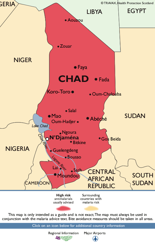
Chad neighbours Libya and Sudan. The risk here, according to the NHS is considered ‘high’ in the entire country. Chad’s border runs along both the northern and southern part of Sudan, but this doesn’t seem to influence the risk factor in northern Sudan much. The last ‘local case’ of ‘malaria’ in Libya was allegedly in 1973.
Morocco is said to be ‘malaria-free’, however, typhoid fever, which appears to have a great deal in common with malaria, is endemic in certain areas, such as Meknes city. According to a study published in 2009 (unfortunately, nothing more recent seems to have been released), typhoid fever “is endemic in the Mediterranean North African countries (Morocco, Algeria, Tunisia, Libya, and Egypt) with an estimated incidence of 10-100 cases per 100,000 persons”.
The countries most affected by malaria are said to be Nigeria, Congo, Uganda, Mozambique and Angola all of which are heavily involved in mining, oil and gas, or combination of both. As discussed in this article, arsenic and lead are contaminants often found near mining areas, but both are also a problem in the oil and gas industry. The aforementioned example of ‘blackwater urine’ was of an individual exposed to arsine gas whilst cleaning a gas tank. A case report from 1950 documents two fatal cases of “Arsine Poisoning in the Smelting and Refining”. Another report published in 1984 documents a case involving five workers who “were exposed to arsine gas after mixing a particularly large quantity of dross with tin ore which was wet because of rain”.
According to ASTM International (American Society for Testing and Materials); “the industrial occurrence of arsine is frequently observed in RNG [renewable natural gas] from landfills and other waste derived sources, and numerous petrochemical, chemical, and metallurgical operations. However, the presence of arsine can go undetected, leading to low level toxic exposure and adverse impacts on fuel performance in various applications”. And in 2022, a literature review discussing arsenic in the petroleum industry notes that “the presence of arsenic in natural gas and liquid hydrocarbons is of great concern for oil companies. In addition to health risks due to its toxicity as well as environmental issues, arsenic is responsible for irreversible poisoning of catalysts and clogging of pipes via the accumulation of As-containing precipitates”.
All in all, there is no shortage of evidence to suggest that various pollutants are the fundamental problem – not the mosquitoes, ticks and other arthropods, and the various microorganisms they carry.
Thank you as always to Caroline Coram for her invaluable help and support in piecing all this information together.





Bravo.
Wow bit of a mind blower, especially the Paris Green. I will probably have to read this a few times to let it fully sink in . Great research!